NiftyReg Boundary Shift Integral Tutorial
Starting from two longitudinal images from the same subject and associated segmentations of region of interest, one can use the Boundary Shift Integral (BSI) to assess the atrophy of the region of interest over time.
The images used in this tutorial are extracted from the MIRIAD database.
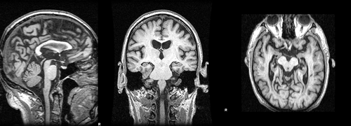
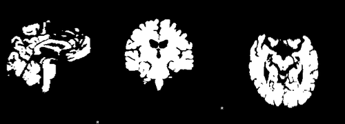
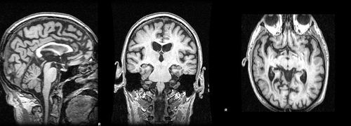
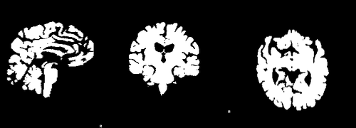
Images 188_A.nii.gz and brain_188_A.nii.gz are a T1-weighted MRI acquired at baseline and its associted full brain segmentation. Images 188_F.nii.gz and brain_188_F.nii.gz are the equivalent images for the follow-up acquisition.
In order to run BSI, we first need to register the baseline and followup image.
reg_aladin -ref 188_A.nii.gz -flo 188_F.nii.gz \
-rmask dil_brain_188_A.nii.gz -fmask dil_brain_188_F.nii.gz \-res ref_A_flo_F_aff_res.nii.gz -aff ref_A_flo_F_aff_mat.txt
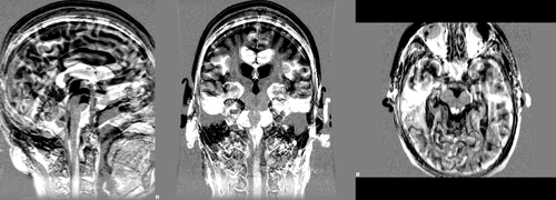
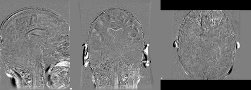
where images dil_brain_188_A.nii.gz and dil_brain_188_F.nii.gz are dilated version of the brain masks of the baseline and followup images respectively. The following images show difference images without registration and with registration respectively
